There have been thousands of injuries & dozens of deaths in Australian children attributable to quad bike use. Warwick Teague, Paediatric Surgeon and Director of Trauma at Melbourne’s Royal Children’s Hospital is passionate about changing this. This is a sobering podcast on a topic that all who work in the Emergency and Trauma scene anywhere, should know about.
Posted in ED Trauma, General Trauma, Paediatric Trauma, PodcastLeave a Comment on ETM Podcast Episode 14 – Warwick Teague – Quad Bikes and Paediatric TraumaCategory: ED Trauma
Tourniquets are back
Largely due evidence from increased military use in conflicts in the last two decades, tourniquet use has re-emerged as an effective way to control haemorrhage from limb injury, usually due to partial or complete amputation or severe crush injury (so called “mangled limbs”) or severe penetrating trauma with major tissue or vascular disruption. Based on reasonable quality data from recent military (and some civilian) studies, tourniquet use for the arrest of life-threatening haemorrhage from exsanguinating limb trauma is now being advocated.
This post provides some general and specific information and practical tips on tourniquet use.
When should I use a tourniquet?
Tourniquets are an effective way to control major haemorrhage from severe limb injury.
Examples of cases where tourniquets may be used include motor vehicle crashes with entrapped/mangled limbs, motorbike crashes with lower limb mangling/amputation, devastating penetrating injuries such as military blast injuries (e.g. land mines, improvised explosive devices), severe crush injuries (e.g.industrial accidents) or deep penetrating injuries such as shark bites. Large, deep incised wounds (e.g. from falls through broken glass or blows from large bladed weapons such as machetes) or firearm injury with major muscle or vascular disruption can also lead to fatal haemorrhage and may benefit from tourniquet use.
TOURNIQUETS SHOULD NOT BE USED FOR SIMPLE PENETRATING WOUNDS, OR HAEMORRHAGE THAT CAN BE CONTROLLED WITH DIRECT COMPRESSION.
Which tourniquet should I use?
Purpose built tourniquets such as the C-A-T tourniquet device or SOF T device (see below) are recommended over improvised devices. Studies comparing different commercially available tourniquets have produced mixed results so no single device can be recommended over another. Simple windlass-style tourniquets are relatively inexpensive, easy to apply and easy to operate.
If a purpose built tourniquet is not available a manual blood pressure cuff (with the tubing clamped) or Bier’s block machine can be used in the emergency department for patients with exsanguinating limb trauma may serve as a temporising measure only as a bridge to more definitive haemorrhage control, however these techniques are not as effective as purpose built tourniquets. Potential dangers of improvised tourniquets include poor fit, inability to control haemorrhage, un-noticed cuff deflation/device failure leading to re-bleeding and difficulty transporting the patient (intra or inter-hospital) with the device in-situ.
General principles of tourniquet use:
Tourniquets should not be used for injuries where haemorrhage control can be achieved by direct pressure.
TOURNIQUETS SHOULD BE TIGHTENED UNTIL LOSS OF THE DISTAL PULSE, OR UNTIL BLEEDING CEASES
There are no clear guidelines on the safe time a tourniquet can be left in place, or to what pressure tourniquets should be inflated. Tourniquet’s left on for too long can cause ischemia to distal tissue, which may lead to cell death or permanent tissue damage and reperfusion of the limb on release of the tourniquet can cause a post-tourniquet reperfusion syndrome characterised by elevated levels of creatinine kinase and lactate, acidosis and hyperkalaemia, as well as distal limb effects including oedema, compartment syndrome, and venous thrombosis, however the absolute risk of these complications is surprisingly low.
Tourniquets also cause compression and ischaemia of the tissues directly under the cuff, which can cause neuropraxia (which may be permanent) and prolonged muscle weakness, although these are also rare complications.
Inflation pressures should not exceed 250-300mmHg in the upper limb, and 300-350mmHg in the lower limb.
Pressures that are high enough to impede venous flow, but not high enough to interrupt arterial flow, will result in continued perfusion of a non-draining limb, leading to oedema, swelling and an increased risk of compartment syndrome.
In elective surgical cases, a maximum tourniquet time of 2-3 hours is usually observed before intermittent release is required. It is important to note that in the emergency trauma setting, mortality and morbidity increases rapidly in patients with vascular limb trauma if tourniquets are left on for more than 45-60 minutes, however this may be more likely related to the overall injury burden sustained leading to tourniquet necessity, rather than due to any potential adverse effects from the tourniquet itself. If there will be a delay of more than an hour to definitive surgical care (for example, delayed retrieval or prolonged transport time), all efforts should be made to obtain vascular haemorrhage control by a surgeon in the referring hospital. If a local surgeon is unavailable, consideration should be given to transporting a surgeon to the patient if this will expedite definitive treatment.
Under no circumstances should periodic loosening of tourniquets be performed without surgical assistance present, as this may lead to uncontrollable re-bleeding.
Patients should not undergo inter-hospital transfer with a tourniquet in-situ if a local surgeon is available to perform temporising surgical control.
Tourniquet use can be very painful and patients will require intravenous analgesia and possibly intubation if pain is uncontrollable.
Practical points for tourniquet use:
- Do not apply tourniquets over clothing – remove all clothing before applying
- Do not apply directly over wounds
- Do not apply over joints – the bony prominences prevent adequate pressure from being applied
- Tighten until bleeding stops/slows or until distal pulse is absent (if distal limb intact/not amputated)
- NB This can be quite painful and the patient may require intubation for pain control/humanitarian reasons
- There is no consensus as to whether more proximal application (i.e. closer to major junctional vessels) or distal application (i.e. closer to wound) is better.
- It is thought that the more proximal the application, the more muscle bulk will be present and less pressure will reach the underlying arteries, hence some providers recommend application as distally as possible (generally 5-10cm proximal to the wound)
- A risk of distal placement is application distal to a missed, more proximal bleeding wound, hence the need for full limb exposure/inspection prior to tourniquet application
- The main determinant of effectiveness of tourniquets is the ratio of device width-to-limb circumference/girth (i.e. the wider the limb, the wider the tourniquet needs to be to be effective)
- Hence, if one tourniquet is ineffective, side-by side (in sequence longitudinally) dual tourniquet use may be effective
- Tourniquet application prior to the onset of shock drastically lowers mortality from haemorrhage compared to application after the onset of shock
- Periodic re-assessment and if required, re-tightening of tourniquets is essential as they can loosen during transport and movement, which can cause re-bleeding and death
- Periodic loosening (e.g. to check for ongoing bleeding) should not be performed until a surgeon is available to address the source of haemorrhage
- There is no defined maximum time for tourniquet application, however it is generally accepted that definitive surgical management should ideally commence within 2 hours of tourniquet application if possible.
- If required to prevent exsanguinating haemorrhage tourniquets can and should be left on until such time as definitive treatment is available
Complications of tourniquets:
Recent battlefield experience in the wars in Middle East have shown a clear risk:benefit ratio and mortality benefit in favour of tourniquet use for exsanguinating limb haemorrhage.
The actual morbidity associated with tourniquets is low
Potential complications of tourniquet use include:
- Ischaemia of distal limb requiring amputation/fasciotomy
- Tourniquet use has not been shown to increase the need for amputation, as the injuries in which tourniquets are used makes it difficult to differentiate between direct or primary limb injury and new or secondary injuries related to tourniquet use.
- In one study approximately 25% of patients with tourniquets applied needed either fasciotomy or amputation, however these were in severely and multiply injured patients with major limb damage.
- Nerve palsies – rare (approx 2% risk)
- Due to peripheral nerve compression
- These are rare and most are transient
- Thrombosis – rare (approx 2%)
- Deep venous thrombosis is a rare complication
- Difficult to distinguish those directly caused by tourniquet versus those occurring as a result of the limb injury
- Skin damage: abrasion, bruising, blisters – rare (approx 1-2%)
- Rhabdomyolysis (rare – approx 1% risk) & acute renal failure (very rare <1% risk)
- Myonecrosis from ischaemic muscle is a rare complication
- Rhabdomyolysis secondary to tourniquet use severe enough to cause acute renal failure is exceptionally rare
Types of tourniquets:
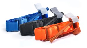
Newer tourniquet devices such as the Combat Application Tourniquet (C-A-T) and SOF Tactical (SOF T) Tourniquet are being used by some pre-hospital services in Australasia.
C-A-T Tourniquet
Image source: Composite Resources
SOF Tactical (SOF T) Tourniquet
Image source: Tacmed Australia
The Australian military has used these and other tourniquets including:
CRoC Junctional Tourniquet – Image not available
JETT: Junctional Emergency Treatment Tool (youtube)
Image source: SAM Medical
If you have any practical tips on tourniquet use or experience you’d like to share, please leave a comment below!
References:
Posted in Circulation, ED Trauma, Haemorrhage control, Trauma Procedures, Trauma Tips, Vascular TraumaLeave a Comment on Tourniquets are back
ETM Interview in Emergency Medicine Australasia
The ETM Course has been growing in reputation and popularity since its launch in November 2013, and what better recognition could we receive than an interview about the course, published in the current edition of EMA (Emergency Medicine Australasia), the main emergency medicine journal for our region. You can find the article here:
http://onlinelibrary.wiley.com/doi/10.1111/1742-6723.12375/full
We’d like to express our thanks to the co-authors (interviewers) Andrew Perry, Joe Rotella, Jenny Jamieson and Rob Mitchell for interviewing Andy about the course, and posing such poignant questions!
It’s so great to have the chance to openly explain the inspiration for, and the the rationale and educational theory behind the ETM Course, and to be able to explain why we think short courses have a place in emergency medicine education.
If you’ve been wondering what the course is about, how it started, and why it’s the most relevant and practical trauma course for emergency medicine, ICU, anaesthetic, surgical and GP trainees and specialists in Australia and New Zealand, check out the article and we hope it inspires you to sign up for a course soon!
Posted in Course News, ED Trauma, General Blog, Trauma EducationLeave a Comment on ETM Interview in Emergency Medicine AustralasiaWill doing the ETM Course help me manage major trauma in the ED?
I can carry on all I like about how good the ETM Course is, and put testimonial videos up from people who’ve just completed it saying how good it is, like this:
httpv://www.youtube.com/watch?v=D6HWJ0BtvNA
And this:
httpv://www.youtube.com/watch?v=4U4fKB4Wmas
But let’s be honest, unless it helps you actually manage a sick, unstable major trauma patient, in a team environment, in your ED, it’s not worth doing. The proof is in the pudding so to speak. This week I received an email from a recent course participant, that I’ve reproduced with permission below, that shows that skills learned on the ETM Course can absolutely be used at your workplace to manage major trauma, and provide you with more confidence in dealing with the group of people who show up to help.
Hi Andy,
I have to tell you about my first Team Leader major trauma experience since the ETM Course.
A quick synopsis:
– 43yo male, high speed motorbike vs power-pole
– intubated at scene, hypotensive, 2xPRBC in transit, collar and pelvic binder
– arrived at ED 1hr after the accident
Things learned from ETM and done well (I thought!!)
– clear role allocation on formation of the “flash team” and shared mental model- “if the FAST is positive straight to OT, activate MTP, clear CT and theatre, all “observers” well out of the way, run through equipment”
– communication including the closed loop- amazing how it drifts away though when the adrenaline kicks in..
– trying to keep out of the way as the team leader- VERY difficult, found myself shuffling round the side of the bed to see various different events so had to remind myself to keep situational awareness and stand back.
– managing seniors! interesting given our discussion about gender roles/ authority gradients etc. By the end of the ED assessment there were 2 surgeons and an anaesthetist there, as well as several registrars – but have to say we were a pretty good team and it went well
End result- Massive transfusion, TXA, 2x thoracostomy and chest drains, positive FAST (unsurprisingly) and fluid in pericardium- spent 21 minutes in ED then went to theatre for damage control. Splenectomy, partial hepatectomy, intra-operative TOE and 22 units of blood products!
I’ve been thinking about it a lot in terms of good lessons learned and things I’d like to improve on – the “Take home message” is the ETM course has given me some really useful tools to manage these situations better– Thank you to you and all your instructors!
So there you have it, a real case from a real course participant. So get over to our registration page, find a date that suits, and come and do the ETM Course!
Posted in ED TraumaLeave a Comment on Will doing the ETM Course help me manage major trauma in the ED?Injury Description in the ED
Trauma means injuries. Gory open wounds that make lay-people faint. Most ED doctors are aware that a “lac is not a lac”, but the entrenched short-hand vernacular we use in ED-speak which is often technically incorrect. Whilst very few medico-legal cases actually hang on whether the open wound you treated was a true laceration or not, or on the opinion of the ED doctor, it is still important that we try to describe injuries we see using the technically correct language. If a case you’ve seen ends up in court, your notes may be subpoenaed, but you will also have to prepare a formal written report after a request from the Police. The more serious the case, the more likely it is that the Police will have already sought the opinion of a forensically trained doctor to see the patient, photograph the injuries, and provide an opinion as to the possible mechanisms of injury. You are within your rights to withhold an opinion about whether the injury is “consistent with the stated cause” if you have not been formally taught how to interpret injuries, which most ED doctors haven’t.
During my ED training I was a Clinical Forensic Medicine Registrar at the Victorian Institute of Forensic Medicine (VIFM) for 6 months, and learned the correct way to describe injuries and compile medico-legal reports, and I’d strongly recommend this job for any ED trainees for your non-ED time, it was an invaulable rotation. For this article however I’m just going to show a few injury examples and their descriptions, so you can see the correct terminology. This is from the upcoming ETM Course manual section on Specific ED Considerations – Injury Description, (Thanks to Dr Nicola Cunningham, Forensic Physician at VIFM for authoring this section).
Bruise:
An area of haemorrhage beneath the skin that is generally due to blunt trauma. Bruises cannot be aged on the basis of their colour, and bruises of identical age and mechanism on the body may not appear at the same time or change at the same rate. The size and shape of a bruise does not necessarily reflect the amount of force or the shape of the object responsible. Some specific bruise subtypes include imprint, fingertip, petechial and periorbital bruises.
Abrasion
A disruption of the outer layers of skin from blunt trauma associated with movement. Abrasions cannot be aged. Scratches and grazes are two specific subtypes of abrasions.
Laceration
A full thickness wound due to blunt trauma resulting in splitting of the skin. The skin usually tears on impact where it overlies a relatively firm surface such as a bony prominence. The wound edges may be ragged, abraded, bruised, inverted. There may be tissue strands bridging the wound.
Incision/Incised Wound
An open wound caused by contact with a sharp object. The wound can occur in any region of the body. The edges are generally regular and clean. There is an absence of tissue bridging strands.
Describing a wound in your notes:
The following characteristics should be documented when describing an open wound in the ED notes:
- Site: The anatomical location
- Size: Approximate length, width +/- depth (if able to assess)
- Shape: linear, crescent shaped, stellate
- Orientation: vertical, horizontal, oblique
- Margins: regular/smooth, irregular/rough, bruised, abraded
- Wound type: laceration, incision, open wound
- Other: direction of track, involvement of deeper structures (tendons, vessels)
If you’re not sure about whether it’s a laceration or an incision – just call it an “open wound” and describe it as above in the notes.
Examples:
An example that implies blunt force:
3 x 1 cm (approx) linear, vertical laceration right lower lip, rough edges, crossing vermillion border:
An example that implies contact with a sharp object:
1 x 7 cm (approx) linear incised wound, flexor aspect mid-right forearm, smooth edges, down to, but not through deep fascia
An example that implies you’re not sure whether blunt or sharp force was applied:
2 x 5 cm (approx) open wound over left elbow, margins smooth but bruised/abraded, olecranon visible at base
Closed wounds can’t be interpreted:
Once a wound has been closed by staples, sutures, glue or steri-strips, or once healing has commenced, no comment can be made about the possible mechanism of injury. You can therefore see how accurate description prior to closure may assist a forensic physician in interpreting your notes (as they may be asked to provide an opinion on such cases).
Photographing injuries in the ED:
When forensic physicians photograph injuries to submit as evidence, certain procedures are followed so as to ensure the evidence is admissible. Firstly, written consent is obtained. Then a color card/white balance shot is taken as the first photo to allow for correct colour grading when the pictures are printed, as colour variation of a wound may be questioned in court. Secondly, the photos must be sequentially numbered with their digital file name, with no interruptions/deletions. This is to ensure that no photo has been removed (which would mean some evidence is missing). The photos are also printed and bound into a booklet by a certified photo processing lab. A digital photo taken on your iphone or ED camera, or taken by the patient, and emailed to the Police will most likely not be admissible as evidence in court.
Assistance with medico-legal report writing:
This subject is beyond the scope of this article, but I wanted to point out that in many states, there will be a Clinical Forensic Medicine (or similarly titled) unit attached to your capital city Coroners office, who will review medico-legal reports for free. This will ensure you won’t trip up on a technicality if you end up having to present your report in court, and I’d recommend you enlist the services of these experts if you are ever required to submit an injury-related report for a court case, to ensure that your injury descriptions are accurate.
Posted in ED Trauma1 Comment on Injury Description in the ED